Decoding Orthopedic Imaging: X rays, MRI, CT SCANS
Welcome
to our in-depth exploration of orthopedic imaging techniques. Orthopedic
imaging plays a pivotal role in the diagnosis and treatment of a wide range of
conditions affecting bones, joints, muscles, and soft tissues. From the classic
X-ray to the advanced MRI and CT scans, understanding these imaging modalities
is essential for diagnosing and treating orthopedic conditions effectively.
X-ray
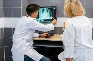
imaging is a vital medical and scientific technique used to create images of
the internal structures of objects, mainly the human body. It utilizes X-rays, a form of
electromagnetic radiation, which can penetrate various materials to different
extents. In medical
applications, X-ray imaging is commonly used for diagnosing fractures,
detecting tumors, examining the chest for pneumonia or lung cancer, and
assessing the condition of internal organs like the heart, lungs, and digestive
system. The process involves the patient being exposed to a controlled
dose of X-rays, with the resulting image providing valuable information to orthopedic
professionals.
Magnetic Resonance Imaging (MRI) is another crucial medical imaging technique that provides detailed images of the internal structures of the body. Unlike X-rays, which use ionizing radiation, MRI relies on a powerful magnetic field and radio waves to generate images. In orthopedics MRI plays a pivotal role in diagnosing a myriad of conditions, from sports injuries and degenerative joint diseases to spinal disorders and soft tissue abnormalities.
Computed Tomography (CT Scanning)
CT
Scans provide high-resolution images of joints, bones, and surrounding soft
tissues offering detailed views of complex anatomy. CT scans provide more
detailed images than conventional X-rays and are particularly useful for
diagnosing and evaluating a wide range of medical conditions. CT scans are fast
and painless, typically taking only a few minutes to complete. However, they do
involve exposure to ionizing radiation, so healthcare providers take precautions
to minimize radiation doses, particularly in children and pregnant women.
Contrast agents, such as iodine-based or barium sulfate solutions, may also be
administered orally or intravenously to enhance the visibility of certain
structures or abnormalities in the images. Overall, CT scanning are valuable
tool in modern medicine for diagnosing and managing a wide range of health
conditions.
Conclusion
Orthopedic
imaging plays a pivotal role in the diagnosis, management, and treatment of
musculoskeletal conditions and injuries. Through various imaging modalities
such as X-rays, MRI, and CT scans, orthopedic practitioners can obtain detailed
insights into the structure and function of bones, joints, muscles, ligaments,
and other soft tissues. Kharghar Multispeciality Hospital in navi Mumbai provides
the integration of advanced imaging technologies with clinical expertise
enabling orthopedic specialists to accurately diagnose orthopedic conditions,
develop tailored treatment plans, and monitor patient progress effectively.
Thank you for visiting here,
Kharghar Multispeciality Hospital.
For more information:
Contact: +91 9594810508 | +91 9594910508
Location: The Crown, first & second floor, plot no 15,16, sector 15, Kharghar, next to D-mart and Reliance Digital, Kharghar, Navi Mumbai, Maharashtra 410210.


Comments
Post a Comment